Acute Ischemic Stroke : Infarct Core Estimation On Ct Angiography Source Images Depends On Ct Angiography Protocol
ADVERTISEMENT
 1
1 2
2 3
3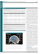 4
4 5
5 6
6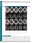 7
7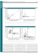 8
8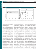 9
9 10
10 11
11 12
12Note: This copy is for your personal, non-commercial use only. To order presentation-ready copies for
distribution to your colleagues or clients, contact us at
Acute Ischemic Stroke :
Infarct
Core Estimation on CT Angiography
Source Images Depends on CT
1
Angiography Protocol
Benjamin Pulli , MD
Purpose:
To test whether the relationship between acute ischemic
Pamela W . Schaefer , MD
infarct size on concurrent computed tomographic (CT)
Reza Hakimelahi , MD
angiography source images and diffusion-weighted (DW)
Zeshan A. Chaudhry , MD
magnetic resonance images is dependent on the parame-
Michael H. Lev , MD
ters of CT angiography acquisition protocols.
Joshua A. Hirsch , MD
This retrospective study had institutional review board ap-
Materials and
R. Gilberto González , MD , PhD
proval, and all records were HIPAA compliant. Data in 100
Methods:
Albert J. Yoo , MD
patients with anterior-circulation acute ischemic stroke and
large vessel occlusion who underwent concurrent CT angi-
ography and DW imaging within 9 hours of symptom onset
were analyzed. Measured areas of hyperintensity at acute
DW imaging were used as the standard of reference for
infarct size. Information regarding lesion volumes and CT
angiography protocol parameters was collected for each pa-
tient. For analysis, patients were divided into two groups on
the basis of CT angiography protocol differences (patients in
group 1 were imaged with the older, slower protocol). Inter-
method agreement for infarct size was evaluated by using the
Wilcoxon signed rank test, as well as by using Spearman cor-
relation and Bland-Altman analysis. Multivariate analysis was
performed to identify predictors of marked ( 20%) overesti-
mation of infarct size on CT angiography source images.
Results:
In group 1 ( n = 35), median hypoattenuation volumes on
CT angiography source images were slightly underestimated
compared with DW imaging hyperintensity volumes (33.0
vs 41.6 mL, P = .01; ratio = 0.83), with high correlation ( r
= 0.91). In group 2 ( n = 65), median volume on CT angi-
ography source images was much larger than that on DW
images (94.8 vs 17.8 mL, P , .0001; ratio = 3.5), with poor
correlation ( r = 0.49). This overestimation on CT angiog-
raphy source images would have inappropriately excluded
from reperfusion therapy 44.4% or 90.3% of patients el-
igible according to DW imaging criteria on the basis of a
100-mL absolute threshold or a 20% or greater mismatch
threshold, respectively. Atrial fi brillation and shorter time
from contrast material injection to image acquisition were
independent predictors of marked ( 20%) infarct size over-
estimation on CT angiography source images.
1
From the Division of Neuroradiology (B.P., P.W.S., R.H.,
CT angiography protocol changes designed to speed imag-
Conclusion:
M.H.L, R.G.G., A.J.Y.) and Interventional Neuroradiology
ing and optimize arterial opacifi cation are associated with
(Z.A.C., J.A.H., A.J.Y.), Massachusetts General Hospital,
Harvard Medical School, 55 Fruit St, Gray 241, Boston, MA
signifi cant overestimation of infarct size on CT angiogra-
02114. Received May 2, 2011; revision requested June 1;
phy source images.
revision received July 27; accepted August 11; fi nal version
accepted August 30. A.J.Y. supported by Neuroradiology
RSNA, 2011
q
Education and Research Foundation/Boston Scientifi c
Fellowship in Cerebrovascular Disease Research. Address
correspondence to A.J.Y. (e -mail: ).
Supplemental material :
/suppl/doi:10.1148/radiol.11110896/-/DC1
q
RSNA, 2011
593
Radiology: Volume 262: Number 2—February 2012
n
ADVERTISEMENT
0 votes
Related Articles
Related forms


 - State Of Connecticut Human Resources")


Related Categories
Parent category: Medical