Acute Ischemic Stroke : Infarct Core Estimation On Ct Angiography Source Images Depends On Ct Angiography Protocol Page 3
ADVERTISEMENT
 1
1 2
2 3
3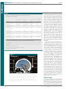 4
4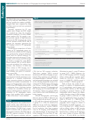 5
5 6
6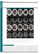 7
7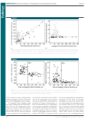 8
8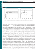 9
9 10
10 11
11 12
12NEURORADIOLOGY:
Infarct Core Estimation on CT Angiography Source Images Depends on Protocol
Pulli et al
Table 1
Materials and Methods
CT Angiography Protocols
This study was approved by our insti-
Parameter
Protocol 1
Protocol 2
tutional review board, and all records
were compliant with the Health Insur-
Time in use
2000–2004
2005–2010
ance Portability and Accountability Act.
CT scanner
LightSpeed Plus, QX/i, or 16
LightSpeed 16 or VCT
We retrospectively examined the clinical
Peak kilovoltage (kV)
140
120
and imaging data in consecutive patients
Tube current (mA)
250
300–800 (Automatic)
with acute ischemic stroke who were
Section thickness (mm)
0.675–2.5
1.25
admitted to our comprehensive stroke
Reconstruction thickness (mm)
5.0
5.0
center between January 2000 and Feb-
Acquisition
From C1 vertebra to vertex; from
From vertex to aortic arch
aortic arch to C1 vertebra
ruary 2010. Inclusion criteria were as
Table speed (mm/sec)
3.75–5.63
9.38–39.38
follows: (a) anterior-circulation acute
Pitch
0.75:1
0.938:1 Or 0.516:1
ischemic stroke; (b) both CT angiogra-
Amount of contrast material (mL)
95–140
65–100
phy and DW imaging performed within
Contrast material injection
3–4
3–4
9 hours of symptom onset and within
rate (mL/sec)
2 hours of each other; (c) large-vessel
Saline chase
None
40 mL at 4 mL/sec
occlusion identifi ed at CT angiography,
Delay (sec)
25 (Fixed); 40 (patients with
Triggered by using SmartPrep with
up to and including third-order (M3)
atrial fi brillation)
region of interest over aortic arch,
middle cerebral artery (MCA) branches;
a threshold of D 50 to 100 HU, and
and (d) absence of reperfusion therapy
a diagnostic delay of 3 seconds
between the CT angiography and DW
imaging sessions. We identifi ed 134
patients who fulfi lled the imaging criteria
and excluded 34 patients (11 patients with
Systems, Milwaukee, Wis). Notably,
eddy current warping and varying the
posterior-circulation strokes [because of
protocol 1 (used from 2000 to 2004)
number of images per section between
streak artifacts potentially compromis-
involved a slower table speed, imaging
28 and 35) were made to the DW im-
ing CT image quality in these regions],
at a fi xed delay following the start of
aging protocol over the study period.
six patients because of severe motion
contrast material administration, and
Ischemic lesions on CT angiography
artifacts at MR imaging, 15 patients
scanning in a caudocranial direction.
source images and DW images were
who had received intravenous tissue
Protocol 2 (used from 2005 to 2010)
outlined independently by two neurora-
plasminogen activator [tPA {Alteplase;
involved a table speed that was up to
diologists (P.W.S [reader 1] and A.J.Y.
Genentech, South San Francisco, Calif}]
10 times faster, SmartPrep triggering
[reader 2], with 18 and 7 years of
between CT angiography and MR imag-
at the aortic arch, and scanning in a
experience, respectively) using dedi-
ing, and two patients with prior infarcts
craniocaudal direction. These changes
cated software (Analyze; Biomedical
in the same territory on the basis of
resulted in imaging the anterior circula-
Imaging Resource, Mayo Foundation,
evaluation of unenhanced CT scans and
tion territory in less than half the time
Rochester, Minn). For CT angiography
their medical records).
after contrast material injection than
source images, window and level settings
with protocol 1 ( Fig 1 ).
were adjusted at the discretion of the
Imaging Protocol and Analysis
For both groups, MR imaging exam-
readers to increase the contrast between
We divided patients into two groups on
inations were performed with a 1.5-T
normal and ischemic brain. Studies were
the basis of changes in the CT angiog-
whole-body unit (Signa; GE Medical
viewed in random order, and readers
raphy protocol that were made at our
Systems). DW imaging was performed
were blinded to all patient information
institution in 2005 (patients in group
by using a single-shot echo-planar spin-
except side of stroke involvement. Infarct
1 were imaged with protocol 1, and
echo sequence. Five images per section
volumes were calculated in milliliters.
2
patients in group 2 were imaged with
were acquired at b = 0 sec/mm
, fol-
CT Angiography Protocol Variables and
protocol 2 [ Table 1 ]). Patients in group
lowed by fi ve images at b = 1000 sec/
Effect on Clinical Management
2
2 were imaged by using the faster CT
mm
in six directions, for a total of 35
angiography protocol. Patients in group
images per section. Imaging param-
Various CT angiography acquisition pa-
1 were imaged with a LightSpeed Plus
eters were as follows: repetition time
rameters ( Table 2 ) were collected and
(four-section), a LightSpeed QX/I (four-
msec/echo time msec, 5000/80–110;
were incorporated into the statistical
section), or a LightSpeed 16 (16-sec-
fi eld of view, 22 cm; matrix, 128 3 128
analysis, including the contrast material
tion) CT scanner; for patients in group
zero-fi lled to 256 3 256; and section
volume and the injection rate, which
2, a LightSpeed 16 or a LightSpeed
thickness, 5 mm with a 1-mm gap. Only
determine the shape of the tissue con-
VCT (64-section) CT scanner was used
minimal changes (introduction of two
centration–time curves. We also calcu-
(all scanners were from GE Medical
180° radiofrequency pulses to minimize
lated the time to imaging of the anterior
595
Radiology: Volume 262: Number 2—February 2012
n
ADVERTISEMENT
0 votes
Related Articles
Related forms


 - State Of Connecticut Human Resources")


Related Categories
Parent category: Medical