Lung Ultrasound Cheat Sheet
ADVERTISEMENT
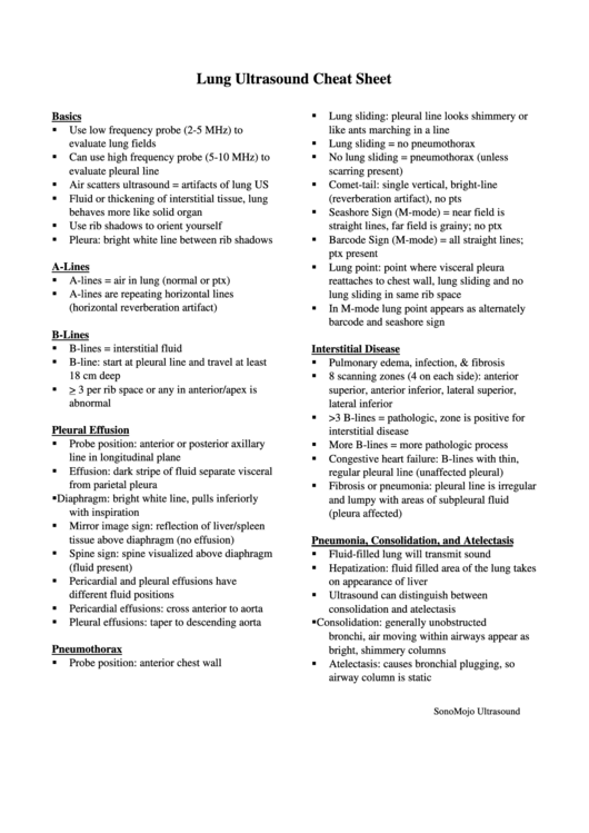
Lung Ultrasound Cheat Sheet
Basics
Lung sliding: pleural line looks shimmery or
Use low frequency probe (2-5 MHz) to
like ants marching in a line
evaluate lung fields
Lung sliding = no pneumothorax
Can use high frequency probe (5-10 MHz) to
No lung sliding = pneumothorax (unless
evaluate pleural line
scarring present)
Air scatters ultrasound = artifacts of lung US
Comet-tail: single vertical, bright-line
Fluid or thickening of interstitial tissue, lung
(reverberation artifact), no pts
behaves more like solid organ
Seashore Sign (M-mode) = near field is
Use rib shadows to orient yourself
straight lines, far field is grainy; no ptx
Pleura: bright white line between rib shadows
Barcode Sign (M-mode) = all straight lines;
ptx present
A-Lines
Lung point: point where visceral pleura
A-lines = air in lung (normal or ptx)
reattaches to chest wall, lung sliding and no
A-lines are repeating horizontal lines
lung sliding in same rib space
(horizontal reverberation artifact)
In M-mode lung point appears as alternately
barcode and seashore sign
B-Lines
B-lines = interstitial fluid
Interstitial Disease
B-line: start at pleural line and travel at least
Pulmonary edema, infection, & fibrosis
18 cm deep
8 scanning zones (4 on each side): anterior
> 3 per rib space or any in anterior/apex is
superior, anterior inferior, lateral superior,
abnormal
lateral inferior
>3 B-lines = pathologic, zone is positive for
Pleural Effusion
interstitial disease
Probe position: anterior or posterior axillary
More B-lines = more pathologic process
line in longitudinal plane
Congestive heart failure: B-lines with thin,
Effusion: dark stripe of fluid separate visceral
regular pleural line (unaffected pleural)
from parietal pleura
Fibrosis or pneumonia: pleural line is irregular
Diaphragm: bright white line, pulls inferiorly
and lumpy with areas of subpleural fluid
with inspiration
(pleura affected)
Mirror image sign: reflection of liver/spleen
tissue above diaphragm (no effusion)
Pneumonia, Consolidation, and Atelectasis
Spine sign: spine visualized above diaphragm
Fluid-filled lung will transmit sound
(fluid present)
Hepatization: fluid filled area of the lung takes
Pericardial and pleural effusions have
on appearance of liver
different fluid positions
Ultrasound can distinguish between
Pericardial effusions: cross anterior to aorta
consolidation and atelectasis
Pleural effusions: taper to descending aorta
Consolidation: generally unobstructed
bronchi, air moving within airways appear as
Pneumothorax
bright, shimmery columns
Probe position: anterior chest wall
Atelectasis: causes bronchial plugging, so
airway column is static
SonoMojo Ultrasound
ADVERTISEMENT
0 votes
Related Articles
Related forms
Related Categories
Parent category: Education
 1
1


