Lab: Blood Exploration Page 3
ADVERTISEMENT
 1
1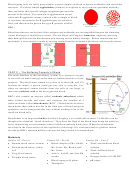 2
2 3
3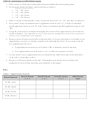 4
4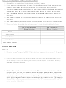 5
5 6
6Procedure
PART A: Histology of Blood Smears
1. Plug in the compound light microscope and turn on the light source. Ensure you are on the
lowest power (shortest objective lens) to begin – this will be a total magnification of 40X.
2. Examine the normal human blood smear slide first. Start by centering the slide in your field of
view and adjusting the coarse adjustment (larger knob of the microscope). This will move the
objective lenses closer/farther away from the stage, bringing the object into rough focus.
3. Next, using the smaller fine adjustment knob, bring your sample into sharp focus. Ensure your
specimen in the center of your field of view.
4. Switch to 100X total magnification WITHOUT touching the coarse adjustment knob. From this
point forward, you can only adjust the fine adjustment. Bring your slide into sharp focus.
5. Carefully switch to 400X total magnification and observe the structure of the normal RBCs
(they will be light pink/red under the microscope). Sketch an RBC in the data table.
6. Have other lab partners observe and sketch the RBCs on the sickle blood smear, frog blood
smear, and dolphin blood smear. Sketch in the data table.
7. Next, identify/find and sketch each of the following types of white blood cells (use your
HANDOUT: Blood to assist in morphology of WBCs. WBCs will be easily identifiable by their
darkly purple-stained nuclei; you can distinguish between the five types based on the shape of
the nucleus and whether or not it is granulated.)
a. Eosinophil: Associated with allergic reactions.
b. Basophil: Associated with allergic reactions.
c. Neutrophil: Protect against pyogenic (pus-causing) microorganisms and participate in
the inflammatory process.
d. Lymphocyte: Includes T-cells and B-cells; generate specific responses that are tailored
to maximally eliminate specific pathogens; “remember” antigens in memory cells.
e. Monocyte: Largest WBCs; replenish macrophages.
8. Compare and contrast the shapes of the WBCs with that of RBCs.
9. Identify/find and sketch platelets.
10. Determine the % composition of your blood smear. Approximately how many RBCs are there
for every WBC? For every platelet?
Data
Normal RBC
Sickle RBC
Frog RBC
Dolphin RBC
Eosinophil
Basophil
Neutrophil
Lymphocyte
Monocyte
Platelet
% Composition of Blood
RBCs?
WBCs?
Platelets?
ADVERTISEMENT
0 votes
Related Articles
Related forms


Related Categories
Parent category: Education