Conformationof 'Eyco(-L-Pro-Gly-)3 And Its Ca2' And Mg2 Complexes Page 3
ADVERTISEMENT
 1
1 2
2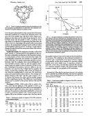 3
3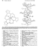 4
4Proc.
Natd.
Acad. Sci. USA
79
(1982)
4521
90
ea
4)
-90
FIG. 3. View of the
peptide
sandwich along the threefold axis with
the
top
peptide ring bonds shown
in
solid black. The octahedral
coor-
dination of the glyc] carbonyls
to
calcium is
seen.
tenon
thus
gives
added stability
to
the unexpected conformation
observed
in
molecule B,
in
which
all
six
carbonyls
point
to
the
same
side of the
peptide
ring,
with
three
of the
oxygens coor-
dinating
to
the
cation
and the other three forming
N-H"O
hydrogen bonds
with the peptide
A
itself.
A
sandwich of this
type
seems
to
be effective
in
sequestering
the
cation
from
the
solvent.
However,
dependingon
the
polarity ofthe surrounding
medium, the intrasandwich hydrogen bonds could be broken
and the peptide sandwich opened, resulting
in
conformational
changes ofmolecule
B
and the
consequent
release of the calcium
ion into
the solvent.
Peptide Mg Complex. The
magnesium
complex,
on
the other
hand, exhibits
a
1:1
stoichiometry for the peptide and the
ion,
the
cation
octahedrally coordinating
to
the three glycyl carbon-
yls of
the
peptide
and
three
water
molecules
(Fig. 5
Left). The
crystallographic
asymmetric unit contains
two
peptide mole-
cules, which show
very
similar coordination
geometry
and
con-
formation.
Both the molecules show noncrystallographic but
close threefold
symmetry
(Table 4),
with
the peptide confor-
mation
being
very
similar
to
that of molecule
A
of the Ca
com-
plex. Puckering of the prolines, however, differs
in
the
two
molecules.
It is
seen
from
Fig. 5
Right that the peptides, the
cations,
and the
water
molecules stack
to
form
an
infinite col-
umn
along
the
crystallographic 21
screw
axis.
The perchlorate
anions
are
located between these columns. The
average
coor-
dination distance of the Mg2+ and the
water
oxygens
is
2.11
A,
whereas that between the
cation
and
the
peptide
oxygens
(2.03 A)
is
significantly
shorter. This difference
in
coordination
distance
may
be indicative of the
greater
basicity of
the
peptide
carbonyl.
Homologues
of (PG)3.
NMR
studies (24)
on
cyclo(-L-Pro-
Gly-)2 show
that
the
backbone of the peptide
is
made
up
of
trans-cis-trasw-cis
peptides like
most
other cyclic
tetrapeptides.
Studies (25)
on
cyclo(-L-Pro-Gly-)4
show that
in
chloroform
it
takes
up a
C4 symmetric
conformation stabilized by
y-turns
and
is made
up
of all trans peptide units. Crystal structure inves-
tigations (26)
on the
rubidium complex show that here also all
Table
3.
Conformation angles (in degrees) of the
(PG)3
molecules
in
the calcium
complex
Mole-
cule
Residue
Xe
Xi
X2
Xs
X4
A
Pro
-64
144 -175 -7
20 -27 -34 -8
Gly
85
179 -177
B
Pro
-68
-24
177
6 -28
39 -34
17
Gly
-84 -157 -170
-90
'(85,
-181)
90
180
4,
degrees
FIG. 4.
4,,
plot of molecule A, molecule
B,
and the 3-y-turn
struc-
ture. Because all three molecules have threefold symmetry the
con-
formation of each molecule
can
be representedjust by
two
points
or one
vector.
The points denoted P and G
represent the
conformations
at
the
proline and
glycine
residues, respectively.
The
coordinates
are
given
in
parentheses.
The
peptide units containing prolines have nearly the
same
orientation with respect
to
the
threefold
axis
in
all
three
cases.
Proline restricts the rotation, but
the 4i
values of molecule A and
molecule B show
a
nearly 1700 difference.
At
glycine residues is
nearly the
same
but 4,values show large differences.
The
conformation
of
the 3-rturn structure
may
be visualized
as
being midway
between
the
structures of molecules A and B.
the peptide bonds
are
trans
but the molecule has deviated
from
C4
symmetry.
The rubidium
ion
has
a
distorted octahedral
en-
vironment
and
is
coordinated
to
four glycyl carbonyl
oxygens
of
one
peptide, another glycyl
carbonyl
of
a
symmetry-related
peptide, and
a
water
oxygen
atom.
A magnesium
complex of
cyclo(-Gly-L-Pro-L-Pro-)2
has also been
reported (27) recently;
it is
a
discrete sandwich complex with approximately twofold
symmetry
relating the
two
peptides of the sandwiches.
We
thank Prof.
EMkan
Blout
for
starting
our
interest
in this
molecule
and Dr. Jake Bello for valuable discussions. This work
was
supported
by U.S. Public Health Service
Grant GM-22490 and
by
the New York
State Department of Health.
Table
4.
Conformation angles (in degrees) of
the (PG)3 molecule
in
the magnesium complex
Mole- Resi-
cule dueno. Residue 4,
4
c
Xo XI
X2
X3
X
A
1
Pro
-63
142 -174
4
9 -18 20-13
2
Gly
79 -171 -173
3
Pro
-57
140
179
9
-9
7 -2
-5
4
Gly
7
7 -173 -179
5
Pro
-64
149 -172 -3
18 -28 25 -13
6
Gly
83
172 -176
B
1
Pro
-63
144-171
3-21
32-29
15
2
Gly
92
172
-175
3
Pro
-59
141
-178 18 -36
40 -30
5
4
Gly
82 -175 -179
5
Pro
-55
139 -178 9 -29 38 -32 15
6
Gly
86
174
177
Average
Pro
-60
143 -176
Average
Gly
84
180 -178
Chemistry:
Kartha
et
aL
ADVERTISEMENT
0 votes
Related Articles
Related forms


 Form Ct-3/4-i - Instructions For Forms Ct-4, Ct-3, And Ct-3-att - General Business Corporation Franchise Tax Returns - 2006
Financial
Form Ct-3/4-i - Instructions For Forms Ct-4, Ct-3, And Ct-3-att - General Business Corporation Franchise Tax Returns - 2006
Financial
 - Exemptions & Schedule 4 - Other Taxes - 2014")
 Form 870-l - Agreement To Assessment And Collection Of Deficiencies In Tax For Partnership Adjustments, Additions To Tax, And Affected Items
Financial
Form 870-l - Agreement To Assessment And Collection Of Deficiencies In Tax For Partnership Adjustments, Additions To Tax, And Affected Items
Financial


Related Categories
Parent category: Miscellaneous