The Muscular System - Anatomy Coloring Sheet Page 17
ADVERTISEMENT
 1
1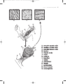 2
2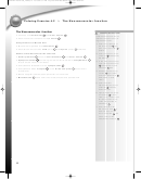 3
3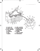 4
4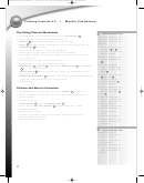 5
5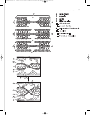 6
6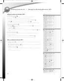 7
7 8
8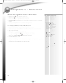 9
9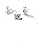 10
10 11
11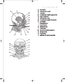 12
12 13
13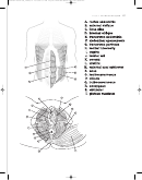 14
14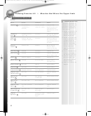 15
15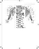 16
16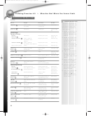 17
17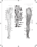 18
18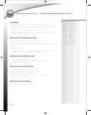 19
19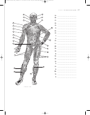 20
20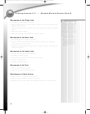 21
21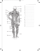 22
22LWBK244-4102G-C04_48-69.qxd 12/12/08 1:22 AM Page 64 Aptara Inc.
Coloring Exercise 4-9
Muscles that Move the Lower Limb
™
F
18, 19,
20
LASHCARDS
AND
COLORING INSTRUCTIONS
Name
Origin
Insertion
Action
Color each muscle and its
name at the same time, us-
Iliopsoas
A
Ilium, lumbar
Femur (lesser
Flexes hip
ing the same color. Color
vertebrae
trochanter)
the anterior and posterior
B
Sartorius
Iliac spine
Tibia body
Flexes thigh,
views together.
leg
1. Review the bones of the
Quadriceps
pelvis and lower limb in
Femoris Group:
Coloring Exercises 3-10
Rectus
Iliac spine
Patella, then tibia
Extends leg;
and 3-11.
femoris
C
flexes hip
2. Review the movements
Vastus lateralis
D
Femur (greater
Patella, then tibia
Extends leg
of the lower limb in Col-
trochanter, linea
oring Exercise 3-13. Re-
aspera)
member that
Vastus medialis
Femur (greater
Patella, then tibia
Extends leg
E
movements at the hip
trochanter, linea aspera)
joint move the thigh, and
Vastus intermedius
Femur
Patella, then tibia
Extends leg
movements at the knee
Adductor
Pubic crest and
Femur (linea
Adducts thigh
joint move the leg
longus
symphysis
aspera)
F
(tibia/fibula).
3. Label some of the bone
Gracilis
Pubis
Tibia
Adducts thigh;
G
features that you see in
flexes leg
this diagram, such as
Adductor magnus
H
Pubis, ischium
Femur (linea aspera)
Adducts thigh
the patella, tibia, and cal-
caneus.
Gluteus medius
I
Ilium
Femur (greater
trochanter)
4. As you read about each
muscle, try to palpate
Gluteus
Iliac crest, sacrum,
Iliotibial tract,
the insertion and origin.
J
maximus
coccyx
femur (linea aspera)
5. Use the muscle to per-
Hamstring Group:
form the action. Use
Biceps
Ischial tuberosity,
Fibula (head) and
Flexes leg;
your fingers to feel the
femoris
K
linea aspera of femur
tibia (lateral condyle)
extends hip
muscle contract.
Semitendinosus
Ischial tuberosity
Proximal tibia
Flexes leg;
L
6. Color the muscle on the
extends hip
diagram. Color the
Semimem-
Ischial tuberosity
Tibia (medial
Flexes leg;
iliotibial tract
a very
T
branosus
condyle)
extends hip
M
light color, because it is
Peroneus
Fibula, tibia (lateral
First tarsal and first
Everts foot
not a muscle.
longus
condyle)
metatarsal of foot
N
st
st
Tibialis
Tibia: lateral condyle/
1
tarsal, 1
Dorsiflexes,
anterior
O
body
metatarsal
inverts foot
Gastrocnemius
P
femur: lateral, medial
Calcaneus (via
Plantar flexes
condyles
Achilles tendon)
foot
Soleus
Fibula (head) and
Calcaneus (via
Plantar flexes
Q
proximal tibia
Achilles tendon)
foot
Extensor digitorum
Tibia
Distal phalanges,
Extends toes
nd
th
longus
R
2
to 5
toes
Flexor digitorum
Posterior tibia
Distal phalanges,
Flexes toes
nd
th
longus
S
2
to 5
toes
Iliotibial tract
Gluteus maximus
Tibia (lateral
Tendon
(tendon)
condyle)
T
64
ADVERTISEMENT
0 votes
Related Articles
Related forms


Related Categories
Parent category: Education