The Muscular System - Anatomy Coloring Sheet Page 3
ADVERTISEMENT
 1
1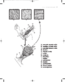 2
2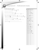 3
3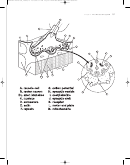 4
4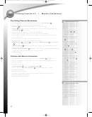 5
5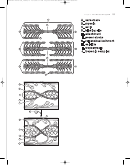 6
6 7
7 8
8 9
9 10
10 11
11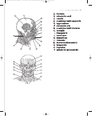 12
12 13
13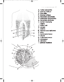 14
14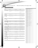 15
15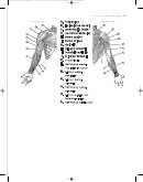 16
16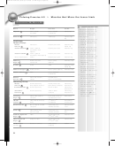 17
17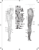 18
18 19
19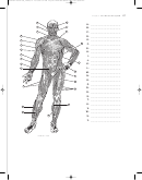 20
20 21
21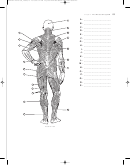 22
22LWBK244-4102G-C04_48-69.qxd 12/11/08 6:11 PM Page 50 LWBK160-3985G-C95_1329-135#C1BC
Coloring Exercise 4-2
The Neuromuscular Junction
The Neuromuscular Junction
COLORING INSTRUCTIONS
• Consists of a muscle cell
and motor neuron
A
B
Color each structure and its
• Each muscle cell contains multiple nuclei
name at the same time, us-
C
ing the same color.
Components of a Muscle Cell
1. Color the cytoplasm of
the muscle cell
light
• Muscle cell organized into sarcomeres
D
A
pink; color the nuclei
C
• Each sarcomere contains actin (thin)
E
and myosin (thick)
F
filaments
purple.
2. Color the column of sar-
Events at the Neuromuscular Junction
comeres
indicated by
D
• Action potential
arrives at axon branches
of a motor neuron
G
B
B1
the bracket.
• Synaptic vesicles
containing stored neurotransmitters (acetylcholine,
)
H
I
3. In the other sarcomeres,
fuse with the neuron membrane
color the actin filaments
• Acetylcholine released into the synaptic cleft
J
red and the myosin
E
filaments
blue.
F
• Acetylcholine binds receptor
K
in the motor end plate
L
(muscle cell
4. Shade the entire motor
membrane)
neuron
light yellow
B
• Bound receptor creates action potential in muscle cell
and the mitochondria
M
• Mitochondria
M
make some neurotransmitters and provide ATP
dark yellow in both
views.
5. Color the arrow
representing the action
potential
travelling
G
down the axon.
6. Lightly shade the synap-
tic vesicles
in both
H
views; use a darker color
for acetylcholine mole-
cules, represented by
small dots
.
I
7 . Use a light color for
the synaptic cleft
, a
J
medium color for the
motor end plate
, and
L
a dark color for the
acetylcholine receptor
.
K
50
ADVERTISEMENT
0 votes
Related Articles
Related forms


Related Categories
Parent category: Education