Acute Ischemic Stroke : Infarct Core Estimation On Ct Angiography Source Images Depends On Ct Angiography Protocol Page 8
ADVERTISEMENT
 1
1 2
2 3
3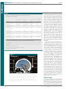 4
4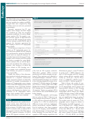 5
5 6
6 7
7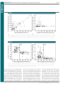 8
8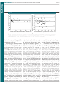 9
9 10
10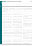 11
11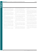 12
12NEURORADIOLOGY:
Infarct Core Estimation on CT Angiography Source Images Depends on Protocol
Pulli et al
Figure 3
Figure 3: (a) Scatterplot shows correlation between infarct volume on CT angiography source images (CTA-SI) and that on DW images (DWI)
for protocol 1 ( ) ( r = 0.912) and protocol 2 ( ) ( r = 0.494). (b) Bland-Altman plot shows agreement between infarct volume on CT angiog-
raphy source images and that on DW images for protocol 1 ( ) (mean, 0.82) and protocol 2 ( ) (mean, 5.0). Dotted line = line of equality.
Figure 4
Figure 4: Graphs show (a) absolute difference and (b) ratio between infarct volume on CT angiography source images (CTA-SI) and that on
DW images (DWI) versus mean time to imaging of the anterior circulation (AC) territory. At imaging times of less than 38 seconds, volume is
overestimated at DW imaging in the majority of cases. Dashed line = line of equality.
of the ischemic territory independently
the basis of a mismatch between infarct
have been inappropriately excluded from
predicted signifi cant lesion overestima-
size on CT angiography source images
treatment on the basis of absolute
tion on CT angiography source images.
and mean transit time abnormality on
thresholds. It needs to be stated, how-
If confi rmed, these fi ndings would
perfusion CT images. In particular, in-
ever, that neither the mismatch concept
have major implications for clinical prac-
farct overestimation on CT angiography
nor absolute infarct size at DW imag-
tices that use CT angiography source
source images could lead to inappropri-
ing is currently recommended outside
images to evaluate brain parenchyma
ate exclusion of patients who may ben-
approved clinical trials as a method of
and estimate infarct core size and for
efi t from treatment. With our current,
selecting patients for treatment. Impor-
clinical trials ( 29–31 ) that are designed
rapid CT angiography protocol, approx-
tantly, many centers are using similar
to select patients for thrombolysis on
imately 45%–60% of patients would
protocols to ours, including protocols
600
Radiology: Volume 262: Number 2—February 2012
n
ADVERTISEMENT
0 votes
Related Articles
Related forms


 - State Of Connecticut Human Resources")


Related Categories
Parent category: Medical